|
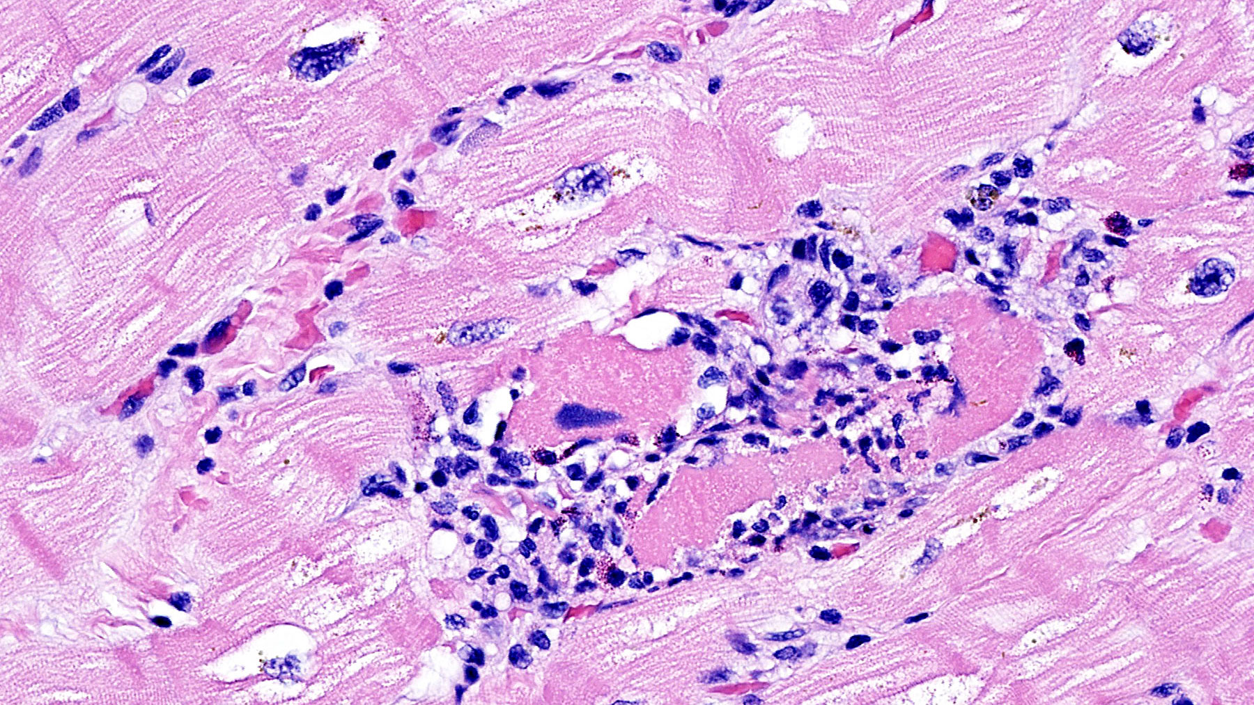
Clinical History: 29 year-old male with history of ulcerative
colitis and drug induced lupus developed acute systolic heart failure.
Histology: H&E stains ( Figures
A, B, C, and D) show Chronic interstitial myocarditis with
mild to moderate infiltration of interstitial spaces with mononuclear
cells admixed with scattered eosinophils and diffuse fibrosis. |