|
Clinical History:
42 year-old male with history of nonischemic
cardiomyopathy with biventricular dysfunction. He had an
Implantable Cardioverter Defibrillator (ICD) for the
past five years, but later developed decompensated heart failure and
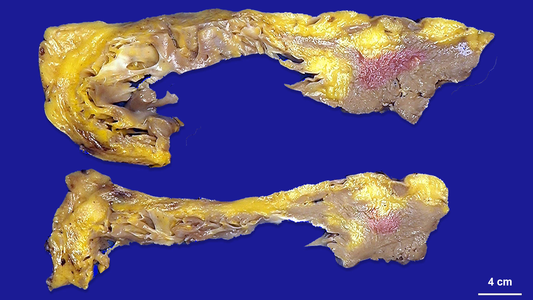
received an orthotopic heart transplant. Sections of the
right and left ventricles from the heart explant (Figure
A) show myocardial walls partially replaced by adipose
tissue (black arrows) that extend from the epicardium
into the myocardium, predominantly in the right
ventricle, but also present in the left ventricle and
septum. Figure B is a close-up of the endocardial
surface of the right ventricle with focal areas of
adipose tissue.
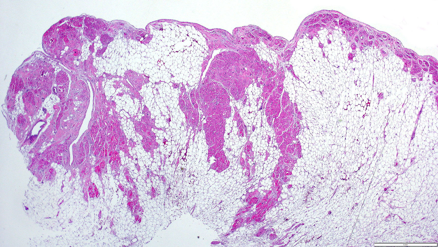
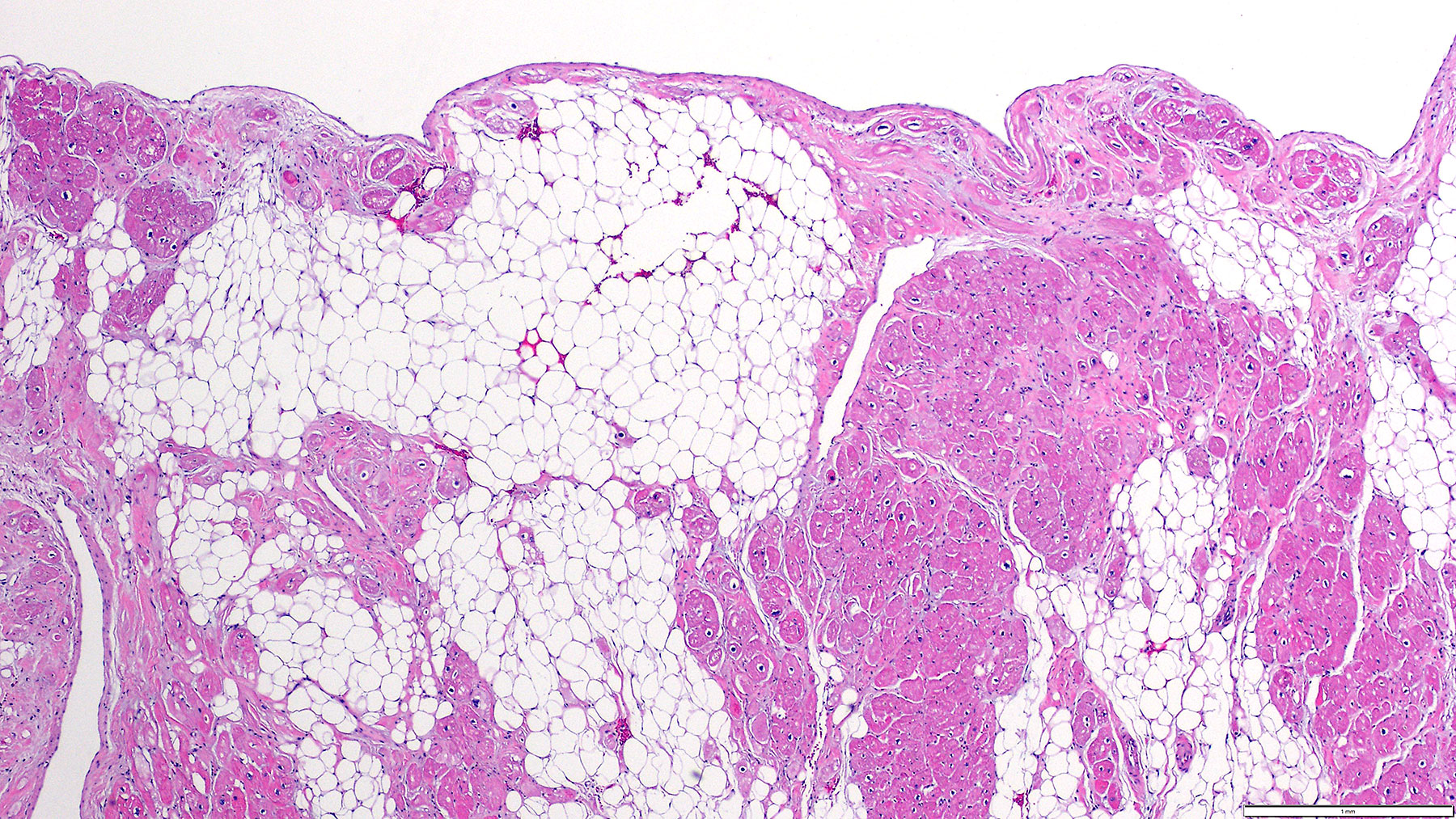
Histology: H&E
staining (Figures C-F) from the explanted heart
shows patchy replacement of the myocardium by adipose
tissue. |